
What is a Cross Bridge cycle? As muscle arrangements, the addition within the thin and thick filaments progress, reducing the time of the sarcomere—the contractile unit of the muscle—using force in the formation of ATP. At the molecular level, this is a cyclic, multistep method that includes required and hydrolysis of ATP, and change of actin by myosin.
When ATP, which is connected to the myosin head, is hydrolyzed to ADP, myosin goes into a high power state attached to actin, forming a Cross-Bridge Cycle When ADP is issued, the myosin head leads to a low power state, leading actin near the middle of the sarcomere. The binding of a new ATP unit separates myosin from actin. When this ATP is hydrolyzed, the myosin top will bind to actin, this time on a lot of actin closer to the end of the sarcomere. Regulatory proteids troponin and tropomyosin, with calcium, work collectively to manage the myosin-actin communication. When troponin binds to calcium, tropomyosin is run away from the myosin-binding position on actin, according to myosin and actin to communicate and muscle compression transpires. This is Cross Bridge
In this article, we at BestComfortBike will give a complete guide on the important topic of What is cross Bridge Cycle. You will be grateful that you have read this article & choice the best Idea for You! Let’s get started!
Table of Contents
You Might Also Like ⇒
- BCB Bicycle Handlebar Grips Fit Many Standard Bikes
- BCB Mountain Bicycle Soft Rubber Grips Fit Many Standard Bikes
- BCB Bicycle Grips For BMX/Road Mountain/Boys And Girls Kids Bikes
- BCB BMX & Mountain Bike Handlebar Grips Non-Slip Soft
- BCB Mountain Bike Handlebar Grips, Two Colour
What is a cross bridge cycle?
In order to understand cross-bridge cycling and its effect in muscle compression, you want to be close with the ‘contractile machine’ that makes muscle compression and the sliding wire system. To briefly recap, muscle compression happens when the thin thread, actin, slides past the thick wire, myosin – This is actually the sliding filament theory. The mechanism that produces the actin filaments to slide through myosin in skeletal tissue, in brief, is as follows:
why is it important in muscle contraction?
learning that a sarcomere is the range of myofibril in two z lines anywhere myosin in middle and an actin fiber is both over and below the myofibril on each front of it, with its central side slightly projecting myosin and its side attached firmly to the z line.
Nervure impulse comes at muscle, making the release of calcium from the sarcoplasmic reticulum of fiber fiber
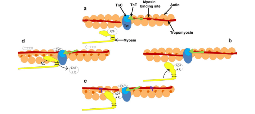
Troponin is attached to tropomyosin which is attached to actin. If the muscle is at ease, tropomyosin is lying in such a place, that it is including the binding positions for myosin heads on actin.
Calcium bind to troponin. This makes it to undergo a conformational switch that pulls the tropomyosin it is connected to out of its comfortable environment, showing binding sites for myosin heads on actin.
Myosin heads covalently attach to the detected required sites.
The myosin head feels a conformational change which pulls the actin along.
It then delivers the actin, reverts to its initial form, reaching out sideways over, and forms a bind with another actin-binding site that is more further along the actin and more like to the z line. This renewed movement is what causes the sliding of the actin fiber through myosin.
Cross bridge cycle refers especially to the action of the cross-bridge, that moving the head and hinge region of the myosin filament. It is really working like a bridge when the end is covalently bonded to actin, and this bridge is continuously staying made and broken as muscle contraction-the cross-bridges are doing cycling, and it is this work that is providing for the wires to start the process they do.
The cross-bridge cycle can be cut down as follows:
Hydrolysis of ATP to ADP and Pi, with results still covalently bonded to myosin, causes it to start an energized state.
Cross bridge connects to actin. It feels like a conformational switch. ADP and Pi are published. You then make an energy stroke (ie cross-bridge moves, pulling actin forward which makes the power stroke (ie the cross-bridge moves pulling the actin along)
ATP binds to myosin, causing the cross bridge to detach.
No 1, Calcium
As control of muscle contraction, calcium absorption is very strictly regulated in muscle fibers. Muscle tissues are in intimate connection with engine neurons. Action potentials in motor neurons announce the neurotransmitter acetylcholine in the region of muscle fibers. This generates an energy potential (depolarization) in the muscle cell, that is provided by the plasma membrane and into invaginations of the plasma membrane described transverse, or T-tubules.
T-tubules run far into the muscle and are next to specific endoplasmic reticulum organelles called the sarcoplasmic reticulum, or SR. Calcium retreated inside the SR is issued when voltage-gated ion channels (ion ways that open and close based on social charges) open in answer to depolarization, releasing calcium ions to enter the cytoplasm, and muscles to contract.
When warning from engine neurons stops, rest of the muscle begins as calcium is pumped back into the SR, reducing the cytoplasmic levels of calcium and providing the SR calcium stores in preparation for the next recession.
No 2, Muscle Degeneration
Healthy muscle can get but infected muscle often loses this capacity. Diseases like myasthenia gravis block motor neuron stimulation of muscle which occurs in muscle atrophy and a reduction in muscle mass. Amyotrophic lateral sclerosis (ALS or Lou Gehrig’s disease) makes motor neurons to degenerate, which then leads to muscle degeneration and atrophy.
Disclaimer
“All brand names and images are Registered Trademarks of their respective companies. All manufacturer’s names, numbers, symbols, and descriptions are used for reference purposes only, and it is not implied that any part listed is the product of these manufacturers or approved by any of these manufacturers.”


